Males and Females Differ in Specific Brain Structures
Medical Xpress, February 11, 2014
Reviewing over 20 years of neuroscience research into sex differences in brain structure, a Cambridge University team has conducted the first meta-analysis of the evidence, published this week in the prestigious journal Neuroscience and Biobehavioral Reviews.
The team, led by doctoral candidate Amber Ruigrok and Professors John Suckling and Simon Baron-Cohen in the Department of Psychiatry, performed a quantitative review of the brain imaging literature testing overall sex differences in total and regional brain volumes. They searched all articles published between 1990 and 2013. A total of 126 articles were included in the study, covering brains from individuals as young as birth to 80 years old.
They found that males on average have larger total brain volumes than women (by 8-13%). On average, males had larger absolute volumes than females in the intracranial space (12%; >14,000 brains), total brain (11%; 2,523 brains), cerebrum (10%; 1,851 brains), grey matter (9%; 7,934 brains), white matter (13%; 7,515 brains), regions filled with cerebrospinal fluid (11.5%; 4,484 brains), and cerebellum (9%; 1,842 brains). Looking more closely, differences in volume between the sexes were located in several regions. These included parts of the limbic system, and the language system.
Specifically, males on average had larger volumes and higher tissue densities in the left amygdala, hippocampus, insular cortex, putamen; higher densities in the right VI lobe of the cerebellum and in the left claustrum; and larger volumes in the bilateral anterior parahippocampal gyri, posterior cingulate gyri, precuneus, temporal poles, and cerebellum, areas in the left posterior and anterior cingulate gyri, and in the right amygdala, hippocampus, and putamen.
By contrast, females on average had higher density in the left frontal pole, and larger volumes in the right frontal pole, inferior and middle frontal gyri, pars triangularis, planum temporale/parietal operculum, anterior cingulate gyrus, insular cortex, and Heschl’s gyrus; bilateral thalami and precuneus; the left parahippocampal gyrus, and lateral occipital cortex.
The results highlight an asymmetric effect of sex on the developing brain. Amber Ruigrok, who carried out the study as part of her PhD, said: “For the first time we can look across the vast literature and confirm that brain size and structure are different in males and females. We should no longer ignore sex in neuroscience research, especially when investigating psychiatric conditions that are more prevalent in either males or females.”
{snip}
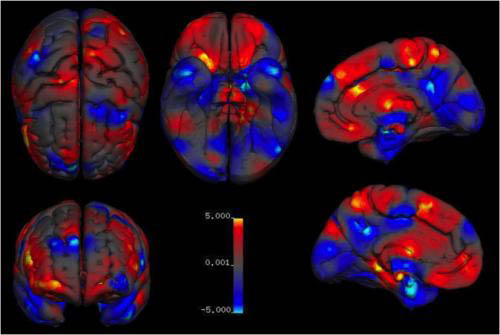
An overview of average regional sex differences in grey matter volume. Areas of larger volumes in women are in red and areas of larger volume in men are in blue.